Anatomy Of Upper Thigh And Hip / Easy Notes On 【Muscles of Anterior Compartment of The ... / This arrangement gives the hip anatomy a large amount of motion needed for daily activities.
byAdmin-
0
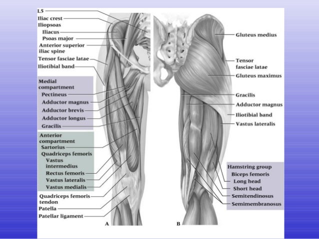
Anatomy Of Upper Thigh And Hip / Easy Notes On 【Muscles of Anterior Compartment of The ... / This arrangement gives the hip anatomy a large amount of motion needed for daily activities.. B, muscles of the anterior thigh compartment. Finally, the hamstring muscles that run down the back of the thigh start on the bottom of the pelvis. Want to learn more about it? Anatomy of the human body. Bones of the lower limb.
Asymmetrical gluteal or thigh skin folds. The uppermost of the medial thigh muscles is the pectineus muscle. Finally, the hamstring muscles that run down the back of the thigh start on the bottom of the pelvis. Knee assessment and hip mechanics online course: Quadriceps, a group of four.
Thigh & Hip from image.slidesharecdn.com Each pelvic girdle consists of a hip bone (coxal bone, innominate bone), which articulates with the head of a femur. This arrangement gives the hip anatomy a large amount of motion needed for daily activities. May 13, 2019 edited by dr. Hip and knee pain and hip and shoulder pain are. The patient lies supine with the hip and knee flexed and the hip externally rotated into the frog leg position. The iliopsoas muscle, which extends from the lower back to upper femur; Hip anatomy, function and common problems. Iliopsoas muscle, a hip flexor muscle that attaches to the upper thigh bone.
It is referred to as a ball and socket joint, and is.
Several muscles cross the front of the hip and create hip flexion, pulling the thigh and trunk toward both muscles cross the floor of the pelvis, emerge at the outer edges of the pubic bones, and finally insert on the inner upper femur (thighbone). Finally, the hamstring muscles that run down the back of the thigh start on the bottom of the pelvis. The upper part of the thigh bone consists of the femoral head, femoral neck, and greater and lesser trochanters. Anatomy hip, thigh and leg muscles. Want to learn more about it? Bends (flexion) the thigh at the hip. The hip adductors refers to a group of five muscles that make up the bulk of the inner thigh mass. Knowing the anatomy of your hip can help you understand the source of any hip pain. This webpage presents the anatomical structures found on hip mri. This webpage presents the anatomical structures found on thigh mri. Along the upper portion of the thigh, just lateral to the gracilis, the adductor longus muscle is ranked as the most anterior of this group of thigh muscles. Foundational anatomy provides medical students with the necessary background in anatomy for success in clerkships. Atlas of human anatomy in cross section.
The paired hip bones are connected. The muscles also require a lot of blood flow, which provides oxygen and nourishment, especially when you're physically active. Hip and knee pain and hip and shoulder pain are. Finally, the hamstring muscles that run down the back of the thigh start on the bottom of the pelvis. Knee assessment and hip mechanics online course:
muscles in the the upper leg | for the thigh where it ... from s-media-cache-ak0.pinimg.com A, anterior and posterior views show the hip joint ligaments. The muscles also require a lot of blood flow, which provides oxygen and nourishment, especially when you're physically active. This webpage presents the anatomical structures found on hip mri. Anatomy hip, thigh and leg muscles. Iliopsoas muscle, a hip flexor muscle that attaches to the upper thigh bone. Local nerves running through and around the pain related to the sacroiliac joints is most commonly experienced in the upper buttock region, usually. The patient lies supine with the hip and knee flexed and the hip externally rotated into the frog leg position. Atlas of human anatomy in cross section.
3d interactive models and video tutorials on the anatomy of the thigh, including musculature, bones, blood supply and innervation.
Atlas of human anatomy in cross section. The cavity of the acetabulum faces obliquely forward, outward, and downward. Jew anatomy atlases, the anatomy atlases logo, and a digital library of anatomy information are all trademarks of michael p. The uppermost of the medial thigh muscles is the pectineus muscle. The hip adductors refers to a group of five muscles that make up the bulk of the inner thigh mass. The paired hip bones are connected. B, muscles of the anterior thigh compartment. The iliopsoas muscle, which extends from the lower back to upper femur; The femur or thigh bone is one of the longest bones in the human body. Local nerves running through and around the pain related to the sacroiliac joints is most commonly experienced in the upper buttock region, usually. Unlike the shoulder girdle, the pelvic girdle is firmly integrated into the axial skeleton: The hip region is located lateral and anterior to the gluteal region, inferior to the iliac crest. In vertebrate anatomy, hip (or coxa in medical terminology) refers to either an anatomical region or a joint.
A, anterior and posterior views show the hip joint ligaments. He also serves the communities of charleston, sc and augusta, ga. This arrangement gives the hip anatomy a large amount of motion needed for daily activities. Bends (flexion) the thigh at the hip. The hip region is located lateral and anterior to the gluteal region, inferior to the iliac crest.
leg | Definition, Bones, Muscles, & Facts | Britannica from cdn.britannica.com Along the upper portion of the thigh, just lateral to the gracilis, the adductor longus muscle is ranked as the most anterior of this group of thigh muscles. The primary function of this group is, surprise, hip these muscles constitute the anatomical classification known as the medial compartment of the thigh. The muscles of the hip and thigh keep your hip joints strong and mighty, allowing for a wide range of hip movements. B, muscles of the anterior thigh compartment. He also serves the communities of charleston, sc and augusta, ga. Knee assessment and hip mechanics online course: Each pelvic girdle consists of a hip bone (coxal bone, innominate bone), which articulates with the head of a femur. Quadriceps, a group of four.
Knowing the anatomy of your hip can help you understand the source of any hip pain.
Related online courses on physioplus. The anterior boundary of the hip adductors is set by. Muscles of the hips and thighs. 431).—at the upper and medial part of the thigh, a little below the medial end of the inguinal ligament, is a large. Iliopsoas muscle, a hip flexor muscle that attaches to the upper thigh bone. Hip movements include flexion, extension, abduction, adduction, circumduction, and hip rotation. Bones of the lower limb. May 13, 2019 edited by dr. Several muscles cross the front of the hip and create hip flexion, pulling the thigh and trunk toward both muscles cross the floor of the pelvis, emerge at the outer edges of the pubic bones, and finally insert on the inner upper femur (thighbone). Finally, the hamstring muscles that run down the back of the thigh start on the bottom of the pelvis. The following nerves serve the gluteal and. Anatomy of the human body. The hip's unique anatomy enables it to be both extremely strong and amazingly flexible, so it can bear weight and allow for a wide range of movement.
Knee assessment and hip mechanics learn how hip and pelvis mechanics can influence the knee powered by physiopedia start course upper thigh anatomy. The following nerves serve the gluteal and.